Patient Education/Resources
The WATCHMAN™ portal is an educational service for use by practicing physicians and allied healthcare professionals. The WATCHMAN portal is not intended for patients or consumers.
This video is for patients who have been prescribed the LifeVest® wearable cardioverter defibrillator (WCD) as well as their families and caregivers. It will tell you how LifeVest works and provide information about assembling, wearing, and caring for the LifeVest WCD.
Electrophysiology Studies (EPS)
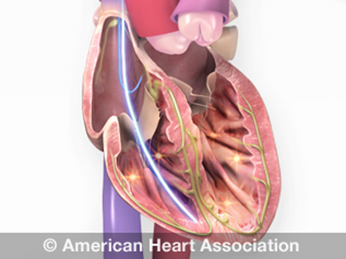
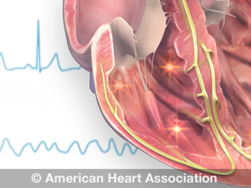
When someone’s heart doesn’t beat normally, doctors use EPS to find out why. Electrical signals usually travel through the heart in a regular pattern. Heart attacks, aging and high blood pressure may cause scarring of the heart. This may cause the heart to beat in an irregular (uneven) pattern. Extra abnormal electrical pathways found in certain congenital heart defects can also cause arrhythmias.
Cardiac catheterization (cardiac cath or heart cath) is a procedure to examine how well your heart is working. A thin, hollow tube called a catheter is inserted into a large blood vessel that leads to your heart.
What to Expect During Cardiac Catheterization
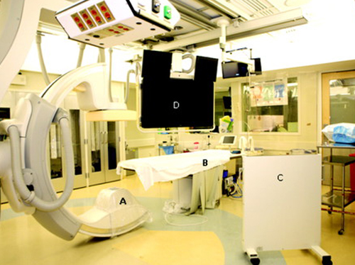
Cardiac catheterization is a test during which flexible tubes called catheters are inserted into the heart via an artery or vein under x-ray guidance to diagnose and sometimes treat certain heart conditions. During right heart catheterization, a vein from the neck, arm, or leg is used to enter the right side of the heart to measure pressures and oxygen content. During left heart catheterization, an artery from the wrist, arm, or leg is used to enter the left side of the heart, usually to perform coronary angiography, which refers to the injection of contrast dye into the coronary arteries to determine the amount of blockage from atherosclerotic plaque.
Cardiac Catheterization (cardiac cath) is a procedure that examines the inside of your heart’s blood vessels using special X-rays called angiograms. Dye visible by X-ray is injected into blood vessels using a thin hollow tube called a catheter.
Implantable Cardioverter Defibrillator (ICD)
ICDs are useful in preventing sudden death in patients with known, sustained ventricular tachycardia or fibrillation. Studies have shown ICDs to have a role in preventing cardiac arrest in high-risk patients who haven’t had, but are at risk for, life-threatening ventricular arrhythmias.
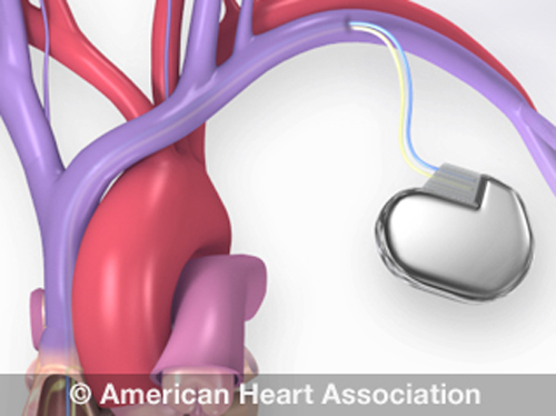
A small battery-operated device that helps the heart beat in a regular rhythm. There are two parts: a generator and wires (leads).
The term “arrhythmia” refers to any change from the normal sequence of electrical impulses. The electrical impulses may happen too fast, too slowly, or erratically – causing the heart to beat too fast, too slowly, or erratically
Catheter ablation is a procedure that uses radiofrequency energy (similar to microwave heat) to destroy a small area of heart tissue that is causing rapid and irregular heartbeats. Destroying this tissue helps restore your heart’s regular rhythm. The procedure is also called radiofrequency ablation.
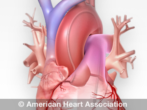
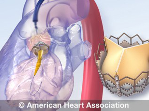
During this minimally invasive procedure a new valve is inserted without removing the old, damaged valve. The new valve is placed inside the diseased valve. The surgery may be called a transcatheter aortic valve replacement (TAVR) or transcatheter aortic valve implantation (TAVI).
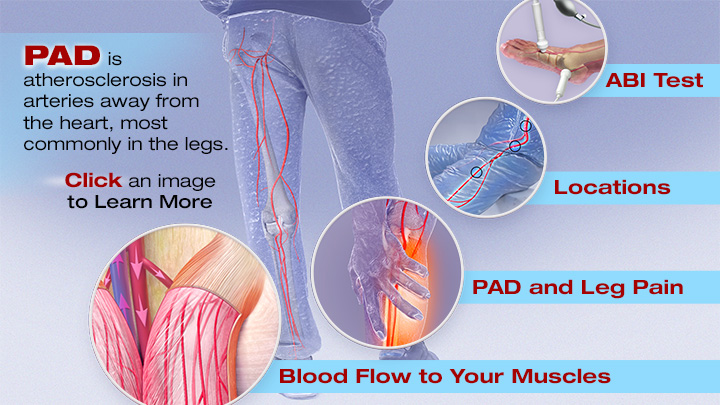
Peripheral Artery Disease (PAD) is atherosclerosis, a buildup of cholesterol and fatty deposits (plaque) which narrows or blocks blood flow to the arteries leading to the arms, legs and feet. The supply of oxygen to cells is also limited due to the plaque buildup in the artery walls.


